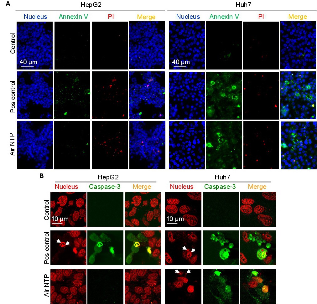
Fig. 7. Microscopy imaging of annexin V and propidium iodide-positive cells after NTP treatment. (A) HepG2 and Huh7 cells were treated with NTP for 60 s, then 6 h after treatment cells were labelled with Hoechst nuclear stain - blue dye, annexin V - green dye and propidium iodide - red dye. Labelled cells were imaged with fluorescence microscopy. Representative images out of three independent experiments are shown. Positive control - 2 µM staurosporine for 4 h. (B) Caspase-3/7 activation assay in Huh7 and HepG2. Cells were stimulated with NTP for 60 s, then 6 h after the treatment cells were labelled CellEvent(tm) Caspase-3/7 Green Assay Kit. Following staining, cells were analysed by confocal microscopy. Representative images out of three independent experiments are shown. NucRed(tm) Live 647 ReadyProbes(tm) Reagent (red dye) was used for nucleus labelling. Positive control - 2 µM staurosporine for 4 h.